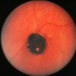
CHRPE represents RPE cells that are twice their normal size and contain densely packed, large melanin granules. CHRPE lesions tend to be unilateral in most cases and can be located anywhere in the retina, primarily temporally or in the periphery. Their diameter varies from approximately 2 to 6 mm, and most commonly appear jet black in color. Although these lesions are usually benign and stable, they should be monitored every 6-12 months because they can enlarge in rare cases.
Unifocal lesions are flat and round with distinct margins. Multifocal ones (aka grouped pigmentation spots or bear tracks) are smaller and tend to aggregate within a confined area. In unifocal lesions, RPE cells lacking melanin form a full or partial halo ring around the lesion, a classical presentation associated with CHRPE. It is possible to see visual field scotomas with large unifocal lesions, but rarely with multifocal ones. This is thought to be due to the defective RPE cells’ inability to phagocytize the photoreceptor’s outer segments, causing the overlying photoreceptors to degenerate.
Another possible finding within the lesion is chorioretinal atrophy, producing one or multiple window defects called lacunae. Fluorescein angiography will show hyperfluorescence through the lacunae, but hypofluorescence in the pigmented area and normal fluorescence in the halo ring.
Differential diagnosis (listed below are features distinguishing the aforementioned condition from CHRPE):
- Choroidal Nevus; indistinct and irregular margins, slate-gray in color, flat to slight elevation, and absent halo, chorioretinal atrophy, or VF scotomas.
- Choroidal Melanoma: almost always greater than 2 mm thick and absent halo.
- Acquired Hyperplasia of the RPE: possible chorioretinal atrophy adjacent to the lesion, irregular margins, absent halo, preceding trauma or inflammatory event and presence of vitreous traction. (Note: hypertrophy means increased size, hyperplasia means reactive proliferation and migration of cells).
- Old chorioretinal scar (i.e. toxoplasmic retinochoroiditis): irregular pigmentation and margins.
Familial Adenomatous Polyposis (FAP) is an autosomal-dominant inherited condition characterized by the development of several intestinal polyps, which can lead to colorectal cancer if left untreated. When FAP is associated with manifestations elsewhere besides the colon, it’s called Gardner’s Syndrome. Although certain ocular findings could occur, CHRPE is the most common. Features that help identify a FAP-associated CHRPE include family history, systemic manifestations, and a bilateral presentation of multiple, non-localized lesions with irregular borders.
Reference: (OptometryStudents.com and the author always recommend a comprehensive review of the sources used in this article)
Jones, William L. Peripheral Ocular Fundus 3rd Edition (pages 18-24).


